The femur, or thigh bone, is the longest and strongest bone in the human body, essential for mobility and weight-bearing. Its robust structure supports various anatomical functions and plays a crucial role in forensic medicine for estimating height, age, and sex.
1.1 Definition and Overview
The femur, or thigh bone, is the longest and strongest bone in the human body, extending from the hip to the knee. It forms the framework of the thigh, connecting the pelvis to the leg. Its sturdy structure includes a spherical head, a narrow neck, and a robust shaft, enabling weight-bearing and movement.
1.2 Importance in Human Anatomy
The femur is the longest and strongest bone, playing a vital role in human anatomy. It supports the body’s weight, facilitates movement, and connects the hip and knee joints. Its robust structure enables mobility, stability, and balance, making it essential for daily activities like walking and running. It also anchors major muscles and ligaments for optimal functionality.
Structure of the Femur
The femur consists of three main parts: the proximal end (upper part), shaft (diaphysis), and distal end (lower part), providing a strong framework for mobility and support.
2.1 Proximal End (Upper Part)
The proximal end of the femur includes the femoral head, a spherical structure, and the femoral neck, a cylindrical segment connecting the head to the shaft. The greater trochanter, a prominent attachment point for muscles, is located on the proximal end, enhancing the bone’s functionality in hip movement and muscle attachment.
2.2 Shaft (Diaphysis)
The shaft, or diaphysis, is the long, cylindrical portion of the femur, providing strength and stability. It is composed of dense cortical bone, making it rigid and capable of supporting heavy loads. The shaft’s surfaces and borders blend smoothly, ensuring efficient weight distribution and muscle attachment, while its slight curvature enhances durability during movement and weight-bearing activities.
2.3 Distal End (Lower Part)
The distal end of the femur, or lower part, includes the condyles and epicondyles. The medial and lateral condyles form the knee joint, with articular surfaces for the tibia and patella. Surrounding these are the epicondyles, which provide attachment points for ligaments and muscles. This region is vital for knee stability, movement, and weight distribution.
Proximal Femur Anatomy
The proximal femur includes the femoral head, neck, and greater trochanter. These structures are critical for hip joint stability, muscle attachment, and facilitating a wide range of motion.
3.1 Femoral Head
The femoral head is a spherical structure at the proximal end of the femur, forming the hip joint with the acetabulum. It is covered by articular cartilage, facilitating smooth movement and weight-bearing; Its blood supply is crucial for its health, and any compromise can lead to conditions like avascular necrosis. It is vital for mobility and stability.
3.2 Femoral Neck
The femoral neck connects the femoral head to the greater trochanter, forming a narrow, cylindrical segment. It plays a critical role in hip joint mobility and is a common site for fractures. Its structure allows for muscle attachments and supports the femur’s weight-bearing function. The neck’s angle and alignment are essential for proper gait and movement patterns.
3.3 Greater Trochanter
The greater trochanter is a large, irregular, quadrilateral eminence on the femur. It serves as an attachment point for several muscles, including the gluteus minimus, gluteus medius, and obturator internus, which are essential for hip abduction and medial rotation. It is a significant anatomical landmark, palpable under the skin when standing, and plays a key role in hip mechanics and stability.
Distal Femur Anatomy
The distal femur forms the lower end of the femur, featuring the medial and lateral condyles. These condyles articulate with the tibia and patella, enabling knee joint stability and movement.
4.1 Lower End of Femur
The lower end of the femur, or distal femur, includes the medial and lateral condyles, which are rounded prominences that articulate with the tibia to form the knee joint. These condyles are covered with articular cartilage, facilitating smooth movement. The intercondylar notch is a groove between the condyles, housing ligaments critical for joint stability and function.
4.2 Condyles (Medial and Lateral)
The medial and lateral condyles are prominent, weight-bearing structures at the distal femur. They articulate with the tibia, forming the knee joint. Articular cartilage covers these surfaces, enabling smooth movement. The medial condyle is larger, while the lateral condyle is more curved. Together, they provide stability and facilitate flexion and extension of the knee.
Functions of the Femur
The femur serves as the primary weight-bearing bone, enabling movement and supporting muscle attachments. Its robust structure ensures stability and facilitates mobility in the human body effectively.
5.1 Weight-Bearing and Support
The femur is the primary weight-bearing bone, distributing forces from the upper body to the lower limbs. Its strong, cylindrical shaft and robust proximal and distal ends provide excellent structural support, enabling the body to maintain posture and engage in activities like standing, walking, and running efficiently.
5.2 Movement and Mobility
The femur facilitates a wide range of movements through its articulation with the hip and knee joints. Its spherical head allows for multi-directional hip movement, while its condyles at the lower end enable flexion, extension, and rotation of the knee. This dual functionality makes the femur essential for walking, running, and maintaining balance.

Blood Supply and Nerve Innervation
The femur receives its blood supply primarily from the femoral artery and its branches. Nerve innervation is provided by the femoral and sciatic nerves, enabling sensory and motor functions.
6.1 Arterial Supply
The femur receives its arterial supply primarily from the femoral artery, which branches into the medial and lateral circumflex femoral arteries. These arteries provide blood to the proximal femur, including the femoral head and neck. Additional supply comes from the perforating branches of the profunda femoris artery, ensuring adequate blood flow to the shaft and distal regions.
6.2 Venous Drainage
The venous drainage of the femur is facilitated by the femoral vein, which accompanies the femoral artery. The medial and lateral circumflex veins drain the proximal regions, while perforating veins along the shaft ensure blood returns to the femoral vein. This network efficiently returns blood from the femur to the heart, maintaining circulatory balance and supporting the bone’s metabolic needs.
6.3 Nerve Supply
The femur receives its nerve supply primarily from the femoral nerve and branches of the sciatic nerve. The femoral nerve innervates the anterior thigh muscles, while the sciatic nerve, derived from the lumbosacral plexus, supplies the posterior thigh and lower limb regions. This extensive network ensures motor control, sensation, and functional mobility of the femur and surrounding tissues.
Common Fractures of the Femur
Femoral neck, shaft, and supracondylar fractures are common, occurring near the hip, mid-thigh, and just above the knee. Causes include osteoporosis, sports injuries, or trauma.
7.1 Femoral Neck Fractures
Femoral neck fractures occur at the neck of the femur, often due to osteoporosis or falls. They are common in older adults and significantly impact mobility. Treatment typically involves surgery, such as plate fixation or arthroplasty, to restore function and alleviate pain. Prompt intervention is crucial to prevent complications like avascular necrosis.
7.2 Shaft Fractures
Femoral shaft fractures occur along the long, cylindrical portion of the femur. These fractures are often caused by high-energy trauma, such as car accidents or falls from heights. They are typically treated with surgical interventions like intramedullary nailing to stabilize the bone and promote proper healing. These fractures can significantly impact mobility and require extensive rehabilitation.
7.3 Supracondylar Fractures
Supracondylar fractures occur just above the knee, involving the lower end of the femur. Common in children, they often result from falls. Treatment may include casting or surgery. These fractures can lead to complications like nerve damage or growth disturbances if not properly managed. Prompt medical attention is crucial for optimal recovery and to prevent long-term issues.
Muscles and Ligaments Associated with the Femur
The femur connects to major muscle groups, including gluteals, psoas, quadriceps, and hamstrings, and is stabilized by ligaments like the iliofemoral and ischiofemoral for hip joint stability.
8.1 Hip Muscles (Gluteals, Psoas)
The gluteal muscles, including the gluteus maximus, medius, and minimus, attach to the femur, facilitating hip extension, abduction, and rotation. The psoas major muscle, part of the iliopsoas, originates from the lumbar spine and inserts on the femoral neck, enabling hip flexion and stabilizing the femur during movement. These muscles are crucial for balanced hip function and mobility.
8.2 Thigh Muscles (Quadriceps, Hamstrings)
The quadriceps muscles, located on the anterior thigh, attach to the femur via tendons, enabling knee extension and stabilization. The hamstrings, comprising the biceps femoris, semitendinosus, and semimembranosus, originate from the ischial tuberosity and insert on the femur and tibia, facilitating knee flexion. Together, these muscles provide balanced movement and stability to the knee joint, essential for walking and running.
8.3 Ligaments (Iliofemoral, Ischiofemoral)
The iliofemoral ligament, the strongest in the human body, connects the ilium to the femur, providing exceptional hip stability. The ischiofemoral ligament, originating from the ischium, reinforces the posterior hip joint. Together, these ligaments enhance joint integrity, prevent dislocation, and facilitate smooth movement, ensuring robust support during dynamic activities like running and climbing.

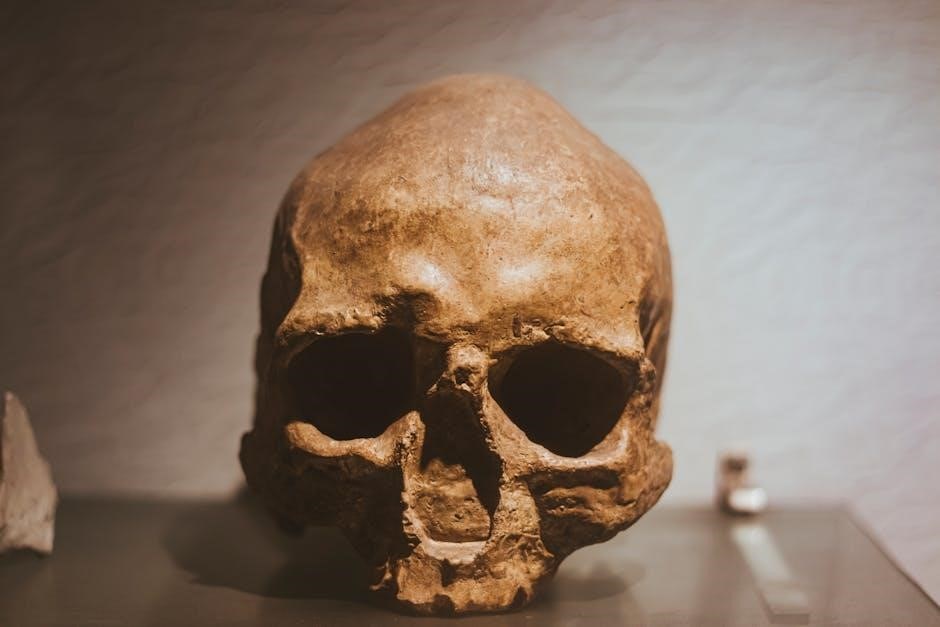
Forensic Importance of the Femur
The femur is crucial in forensic anthropology for estimating height, sex, and age due to its size and durability, aiding in identifying human remains and solving criminal cases.
9.1 Estimation of Height and Sex
The femur is a valuable tool in forensic anthropology for estimating height and sex. Its length correlates strongly with overall height, while sexual dimorphism in size and robustness helps determine sex. The femoral head diameter and bone density are key indicators, aiding in accurate identification of remains and reconstructing biological profiles in criminal investigations.
9.2 Determination of Age
The femur aids in age determination through analysis of bone density, cortical thickness, and epiphyseal union. Radiographic and histological studies reveal age-related changes, such as osteon remodeling and Haversian canal size. These factors help forensic anthropologists estimate age by examining bone structure and degenerative patterns, providing critical insights in identifying human remains and solving forensic cases accurately.
Related Anatomy
The femur connects with the pelvis at the hip joint and the tibia and patella at the knee, forming essential joints for movement and stability, crucial for locomotion and weight distribution.
10.1 Hip Joint (Acetabulum and Femoral Head)
The hip joint is a ball-and-socket joint formed by the femoral head and the acetabulum of the pelvis. The femoral head, a spherical structure at the femur’s proximal end, articulates with the acetabulum, enabling omnidirectional movement. This joint is stabilized by strong ligaments, facilitating activities like walking, running, and maintaining posture, while distributing weight effectively.
10.2 Knee Joint (Femur, Tibia, and Patella)
The knee joint, a hinge-type synovial joint, connects the femur, tibia, and patella. The femur’s distal condyles articulate with the tibia, while the patella glides in the femoral groove. This joint enables flexion and extension, essential for walking, running, and climbing. Ligaments and surrounding muscles provide stability, making it a critical structure for lower limb mobility and weight distribution.
Diagrams and Illustrations
This section includes detailed 3D renderings, front and side views, and cross-sectional illustrations of the femur. These visuals provide clear, labeled representations for educational and clinical reference.
11.1 Front and Side Views
Front and side views of the femur provide detailed visual representations of its anatomy. These illustrations highlight the femur’s elongated shaft, rounded head, and condyles. Labeled diagrams emphasize key features such as the femoral neck, greater trochanter, and distal condyles, offering clarity for educational and clinical purposes. These visuals aid in understanding the bone’s structure and spatial orientation.
11.2 Cross-Sectional Views
Cross-sectional views of the femur reveal its internal structure, showcasing the cortical bone’s thickness and the medullary cavity. These views are essential for understanding the bone’s composition, density, and growth patterns. They also highlight the femur’s structural adaptations for weight-bearing and stress distribution, making them invaluable for anatomical studies and clinical analyses of bone health.
Clinical Significance
The femur’s strength and structure are vital for weight-bearing and mobility. Its susceptibility to fractures, particularly in conditions like osteoporosis, highlights its clinical importance in orthopedic and forensic medicine.
12.1 Osteoporosis and Fracture Risk
Osteoporosis significantly increases the risk of femur fractures, particularly in the femoral neck. This condition weakens bone density, making the femur more susceptible to breaks during falls or minor trauma.
Fractures in the femur, especially in older adults, can lead to severe mobility issues and a reduced quality of life. Early detection and treatment of osteoporosis are critical to preventing such injuries and maintaining bone health.
12.2 Femur Injuries in Sports
Femur injuries in sports are often severe due to high-impact trauma. Common injuries include fractures from direct blows or extreme stress, and overuse stress fractures in endurance sports. These injuries can significantly impact an athlete’s career, requiring prolonged recovery and rehabilitation. Proper preventive measures and timely medical intervention are crucial to mitigate long-term damage.
The femur, as the longest and strongest bone, plays a vital role in human anatomy, mobility, and forensic science. Understanding its structure and functions remains essential for medical advancements and anatomical studies.
13.1 Summary of Key Points
The femur, as the longest and strongest bone, plays a critical role in human anatomy, supporting movement and weight-bearing. Its structure, including the head, neck, shaft, and condyles, facilitates joint connections and muscle attachments. Its clinical significance in fractures, osteoporosis, and forensic applications underscores its importance in medical and anatomical studies.
13.2 Future Research Directions
Future research on the femur should focus on advancing biomechanical studies, improving fracture treatment methods, and exploring regenerative therapies. Investigating gender and age-related anatomical variations could enhance forensic applications. Additionally, developing personalized surgical approaches and preventive strategies for osteoporosis-related fractures remains a priority for improving patient outcomes and quality of life.